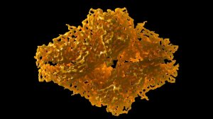
The 2017 Nobel Prize in Chemistry has been awarded to three scientists whose work revolutionized the way researchers visualize the molecular machinery of life. Jacques Dubochet, Joachim Frank, and Richard Henderson were recognized for their development of cryo-electron microscopy, a technique that has made it possible to capture three-dimensional images of biomolecules at atomic resolution.
The technology, often abbreviated as cryo-EM, allows scientists to freeze biological samples in their natural state and observe them under an electron microscope without damaging their structure. This breakthrough has filled a critical gap left by earlier imaging methods, enabling the study of processes that were previously invisible to researchers. By freezing biomolecules mid-movement, scientists can now witness transient states and dynamic interactions that are essential to understanding the chemistry of living organisms and developing new pharmaceuticals.
For decades, the use of electron microscopes on living cells was considered impossible because the electron beams could destroy biological material. Henderson, a researcher at the MRC Laboratory of Molecular Biology in Cambridge, UK, overcame this obstacle in 1990 when he successfully produced a three-dimensional image of a protein at atomic resolution using an electron microscope. His achievement demonstrated that high-resolution imaging of biomolecules was feasible, paving the way for the technique's broader application.
Frank, a professor at Columbia University, contributed a key computational method between 1975 and 1986. He developed a way to combine multiple two-dimensional images, each fuzzy and noisy, into a single sharp three-dimensional model. This algorithmic approach is fundamental to the cryo-EM workflow, allowing researchers to reconstruct detailed structures from many noisy observations.
Dubochet, who was affiliated with the University of Lausanne in Switzerland, solved a different problem: how to prepare biological samples for imaging without destroying them. He devised a method of rapidly cooling water so that it solidifies into a glass-like state, rather than forming ice crystals that would damage the sample. By adding water to the microscope's vacuum and cooling it quickly, he ensured that biomolecules retained their natural shape during imaging.
Since these initial breakthroughs in the 1970s and 1980s, the three laureates have continued to refine the technique. It was not until 2013 that cryo-EM achieved the desired atomic resolution, marking a turning point in its utility. Since then, the technology has produced never-before-seen images of a wide range of biomolecules, from the Zika virus to proteins that confer antibiotic resistance.
Henderson, speaking to Nobel Media, described the impact of cryo-EM on structural biology. He noted that the technique has opened up a previously unapproachable area of the field, particularly for structures that were resistant to other methods such as X-ray crystallography or nuclear magnetic resonance spectroscopy. His comments underscore the significance of the award, which recognizes not just a technical achievement but a fundamental shift in how scientists can explore the molecular basis of life.
The Nobel Committee's choice highlights the practical benefits of cryo-EM, which has become an indispensable tool in drug development and basic research. By enabling the visualization of proteins and viruses in their native state, the technology offers insights that could lead to new treatments for a variety of diseases.